
Okayama University Develops Innovative Imaging Technique to Map Lipid Distribution in C. elegans


Okayama University has recently made significant strides in the field of biochemical imaging with the introduction of a novel mass spectrometry imaging method designed to visualize lipid distribution within the organism Caenorhabditis elegans, more commonly known as C. elegans. This research, spearheaded by Professor Masazumi Fujiwara of Okayama University's Graduate School of Natural Science and Technology, alongside a dynamic team including students and faculty from Konan University, has opened a new frontier in understanding lipid metabolism and related diseases.
The main achievement of this collaborative effort is the ability to create successive tissue slices of C. elegans while preserving its internal structure, allowing for a three-dimensional visualization of lipids. At a remarkable scale of 10µm, each slice can be analyzed for its lipid composition, granting researchers unprecedented insights into the spatial distribution of lipids across different organs. This detail is vital for studying complex biological processes, as lipids play crucial roles in metabolism and cellular functions.
In this research, advanced mass spectrometry techniques were combined with traditional staining methods to effectively map lipid distribution. This integration not only enhances the accuracy of the imaging but also enables a dual verification approach—researchers can validate their mass spectrometry data using information obtained from staining, thereby confirming the lipid profiles observed in the slices.
C. elegans serves as a valuable model organism due to its genetic similarities with mammals—which include critical pathways involved in lipid metabolism. This similarity makes it an ideal subject for studying various human disorders such as obesity, diabetes, and neurodegenerative diseases. By applying this new imaging technique, researchers expect to track lipid dynamics at the individual organism level, paving the way for early diagnostics and the discovery of new biomarkers crucial for drug development.
The implications of this research extend beyond mere academic interest. The technological advancements achieved through this study can be significant for pharmaceutical industries looking to develop more effective treatments for lipid-related diseases. Understanding lipid distribution and dynamics can lead to better-targeted therapies and innovative drug designs. Furthermore, this method holds the potential to analyze how factors such as aging, stress, and nutrition impact lipid metabolism, thereby enriching fundamental research in biological sciences.
Professor Fujiwara expressed excitement about the challenges overcome in developing this imaging technology. The precision required for creating frozen micro-slices of C. elegans is high, and the successful construction of three-dimensional images from these slices was a breakthrough that surpassed initial expectations.
Sara Mandić, a graduate student involved in this research, echoed these sentiments, emphasizing the joy of achieving successful imaging for the first time with this new method. She looks forward to exploring further applications of this innovative technique in future research endeavors.
The research results were published in the journal Scientific Reports on July 9, 2025, opening new avenues for molecular visualization and quantitative analysis in model organisms. Researchers are hopeful that this methodology will contribute to a broader understanding of lipid dynamics and ultimately lead to significant advancements in healthcare, drug discovery, and environmental science. The excitement surrounding this project underscores the potential of collaborative academic research to drive innovation and improve human health.
This study not only highlights the capabilities of Okayama University in pushing the boundaries of scientific research but also exemplifies the importance of interdisciplinary collaboration in tackling complex biological questions. As the field of imaging technology progresses, this method could become a standard in lipid research across various biological systems, promising to enhance our understanding of metabolism in health and disease.
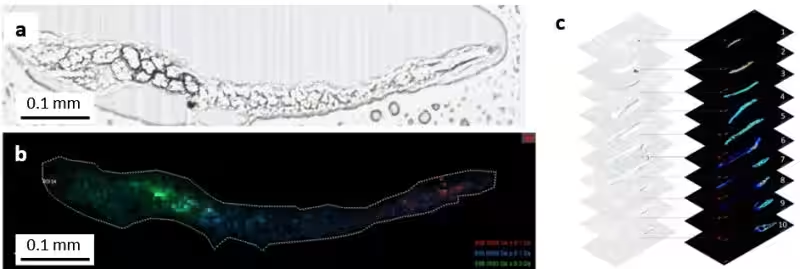
Topics Health)

【About Using Articles】
You can freely use the title and article content by linking to the page where the article is posted.
※ Images cannot be used.
【About Links】
Links are free to use.