
Technology to Visualize the Internal Dynamics of Microplastics Accelerates Risk Assessment in Real Environments


Technology to Visualize Microplastics in the Body
Recent research from the Tokyo University of Science has introduced an innovative method for tracking the internal behavior of microplastics (MPs) in living organisms. This breakthrough is achieved through the development of fluorescent microplastic particles that incorporate near-infrared dyes. The collaborative study led by Associate Professor Masakazu Umezawa and Professor Kohei Soga, alongside graduate students, aims to provide insights into the health impacts of microplastics, which continue to permeate our environment and bodies without a clear understanding of their implications.
Understanding the Need
Microplastics, made from materials such as polypropylene (PP), polyethylene (PE), and polystyrene (PS), have infiltrated every corner of our planet—from oceans to soil, and even our bloodstream. As they enter our bodies through food and water, their dynamics within biological systems remain largely uncharted territory. Traditional tracking methods typically require organ extraction, limiting our ability to observe continuous in vivo processes.
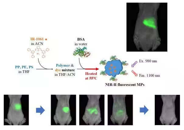
To overcome these limitations, this research applies near-infrared (NIR) imaging, which allows for non-invasive monitoring of particles within living organisms. By synthesizing fluorescent MP particles from multiple plastic materials, researchers have successfully demonstrated the ability to trace their movement within the mouse model.
Key Findings
The study's methodology involved using NIR fluorescent dye, IR-1061, known for its properties that facilitate deep imaging, to create fluorescent MPs. The team was able to develop tracking capabilities that extended beyond just PET materials, successfully including other plastics like PP, PE, and PS. Through this innovation, they examined the absorption of orally ingested MPs in mice, ultimately revealing that a minuscule percentage of these particles are actually absorbed by the body, necessitating data-driven discussions regarding health risks associated with MPs.
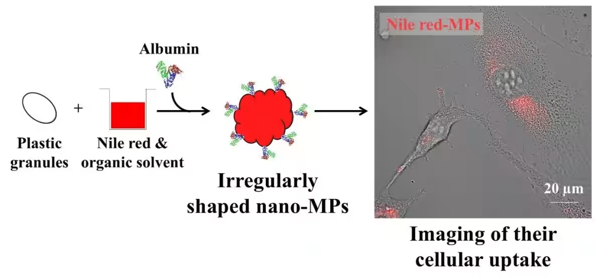
Moreover, the team explored irregularly shaped MP particles—a representation of real-world microplastics—using albumin as a surfactant. This effort was aimed at understanding how shape influences cellular uptake and toxicity, with findings showing that even at a low concentration, irregular MPs demonstrated higher cellular uptake compared to spherical ones.
Implications for Future Research
The study raises new concerns that must be addressed, particularly the suggestion that irregularly shaped MPs might pose greater toxicity compared to their spherical counterparts. This insight challenges the status quo of previous research that primarily relied on spherical MPs, indicating a need for further investigation into real-world particulate forms and their risks. The research concluded with the expectation that further studies would build on these findings, offering a comprehensive risk assessment framework concerning microplastics.
Conclusion
Highlighting the global plastic waste crisis that is projected to escalate significantly by 2040, this research stands as a crucial step toward understanding the intricate dynamics of microplastics in biological systems. The technologies developed not only allow for the tracking of MPs within live specimens but significantly contribute to the growing body of knowledge required to assess and mitigate the potential health risks associated with these persistent pollutants. Associate Professor Umezawa emphasizes the importance of bridging scientific research with environmental realities to foster a deeper understanding of the ramifications of plastic pollution.
This groundbreaking research was funded by the Japan Society for the Promotion of Science (JSPS) Grants-in-Aid and will be elaborated on in upcoming publications in Environmental Science: Advances.
Topics Health)
【About Using Articles】
You can freely use the title and article content by linking to the page where the article is posted.
※ Images cannot be used.
【About Links】
Links are free to use.