


New Insights into Autism Spectrum Disorder Through Neural Representation of Body Parts
Unveiling the Neural Representation of Body Parts in Adults with Autism Spectrum Disorder
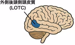
A recent research study led by Yuto Kurihara, a junior associate professor at Waseda University, along with Rieko Oosu, a professor, and Yuuko Okamoto, a visiting senior researcher, has revealed important insights into how adults with Autism Spectrum Disorder (ASD) perceive the bodies of others. This examination utilized functional magnetic resonance imaging (fMRI) to visualize brain activity while focusing on specific body parts, particularly through the lateral occipitotemporal cortex (LOTC).
Key Findings
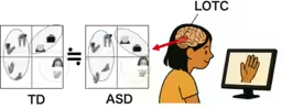
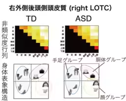
The significant finding of this study is that both adults with ASD and those with typical development (TD) exhibit similar patterns of brain response when observing body parts. The researchers conducted a representational similarity analysis (RSA), which categorizes the brain's activity patterns into three main clusters: "face," "limbs," and "torso." This suggests that the fundamental visual processing of body parts is comparable between the two groups, indicating that the challenges faced by ASD individuals in interpersonal communication might not stem from the way they see bodies but rather from the interpretation and understanding of visual stimuli at a higher cognitive level.
Background of the Research
Previous studies highlighted that individuals with ASD often struggle to interpret emotions and intentions from the bodies and faces of others. It was believed that their difficulties in communication were linked to visual processing or social interaction challenges. Existing brain imaging studies indicated weaker responses in specific brain regions associated with body and facial recognition, namely the Extrastriate Body Area (EBA) and the Fusiform Face Area (FFA), as compared to TD individuals. However, the nuances of how bodies are visually perceived—how different body parts are distinguished—had not been thoroughly investigated until now.
Research Methodology
The primary goal of this study was to elucidate how adults with ASD categorize body parts in their brains when viewing them. Unlike many previous studies that focused on the overall strength of brain responses, this research sought to compare how the different body parts are semantically organized in the brains of both ASD and TD groups. To achieve this, a series of images depicting various body parts (including the face, hands, arms, legs, chest, and waist) were shown to participants, while their LOTC activity was recorded using fMRI technology.
After obtaining brain activity data, the study employed the RSA methodology to examine the similarities between the brain responses to different stimuli. By focusing on the spatial patterns of brain activity in response to various stimuli, it clarified how the brain treats different stimuli as "close" or related.
Results and Implications
The analysis yielded compelling results, revealing that both ASD and TD individuals categorize body parts in a similar structural manner within the LOTC. The similar patterns showed that when observing body parts, both groups organized them into the same three meaningful categories: "face," "limbs," and "torso." Furthermore, this finding was statistically significant and supported by another classification method called multi-voxel pattern analysis (MVPA), which confirmed that these categories were distinctly recognized by both groups. This indicates that the cognitive processes involved in visual categorization of body parts are very similar for ASD individuals as well as their TD counterparts.
Societal Impact and Future Directions
The findings suggest that the visual representation structure for body parts is not fundamentally different in individuals with ASD when compared to those with typical development. This challenges the notion that the visual aspect of body perception is the root cause of communication difficulties typically associated with ASD. Furthermore, the absence of a significant correlation between LOTC activity and the features of ASD, such as communication challenges and sensory sensitivities, indicates that these issues may be attributed to differences in higher cognitive functions rather than visual perception.
Despite the groundbreaking nature of this research, it is worth noting that the study focused solely on adults with ASD. Thus, further investigation involving children and adolescents with ASD is necessary to better understand how visual representations and associated social difficulties evolve over time.
Conclusion
This study emphasizes the importance of exploring the neural foundations of body perception in individuals with ASD. By identifying that the initial visual recognition abilities of adults with ASD are structurally similar to those of typical individuals, it strengthens the argument that effective intervention and support mechanisms must address cognitive processing rather than solely focusing on visual perception. Ultimately, these insights might contribute to constructing a more inclusive society for individuals with ASD.
Topics Health)


【About Using Articles】
You can freely use the title and article content by linking to the page where the article is posted.
※ Images cannot be used.
【About Links】
Links are free to use.