Revolutionizing Fluorescent Microscopy with øCAO Technology for Enhanced Cellular Imaging


Exciting Advances in Fluorescent Microscopy
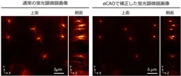
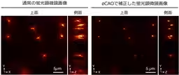
Recent advancements in fluorescent microscopy have opened new avenues for cellular imaging, making it possible to view the intricate internal structures of living cells with unprecedented clarity. A team of researchers from the National Institute of Information and Communications Technology (NICT), Kyoto University, and Utsunomiya University has developed a pioneering computational technique named "øCAO" (phi Computational Adaptive Optics) that automatically corrects for optical distortions in images captured through fluorescent microscopes.
The Challenge of Optical Distortion
One of the significant challenges in fluorescent microscopy is the optical distortion caused by variations in how light passes through different areas within living cells. This distortion often results in blurry or dark images, making it challenging for researchers to observe the true structure of the cells. The newly developed øCAO technique employs computational methods similar to those used in astronomical optics to automatically enhance the clarity of images without requiring expensive modifications to existing equipment.
Innovative Approach: øCAO
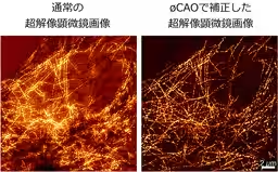
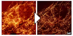
The core advancement of øCAO lies in its ability to process and correct the optical distortion using mathematical algorithms. These algorithms allow the system to refocus images post-capture, enhancing the visibility of cellular structures and deeper tissue layers. Not only does this technique work with standard fluorescent microscopes, but it is also applicable to super-resolution microscopy, significantly boosting the observational capabilities in life sciences.
Since its conception, øCAO has demonstrated the ability to transform indistinct images into clear representations, revealing structures that were previously obscured due to optical disturbances. The research group discovered that by applying computational techniques, they could restore the images to their original clarity without needing supplementary hardware additions, a significant leap forward for researchers.
Application and Impact on Life Sciences
This technology has vast implications for the fields of medical research and drug discovery. By improving clarity and resolution in fluorescent microscopy, researchers can gain better insights into diseases and cellular abnormalities. The potential for better diagnosing diseases and accelerating drug development processes has made this innovation a crucial asset in biomedical research.
The results of this study were published in the peer-reviewed journal "Communications Engineering" on March 9, 2026, underscoring the scientific community's recognition of this breakthrough.
The Path Forward
Looking ahead, the research team intends to broaden the application of øCAO to different super-resolution microscopy techniques and two-photon microscopy, which will allow further exploration of cellular structures at even greater depths. This will enhance the accuracy of the sensing technology that reads biological information and support more intricate foundational and applied research in biology.
The demo program for the øCAO is available online, allowing other researchers to explore this innovative method. The collaboration between NICT and universities showcases the integration of various scientific disciplines to push the boundaries of understanding in the life sciences field.
Through this remarkable advancement, øCAO not only elevates the existing capabilities of fluorescent microscopy but also contributes to the efficiency of research efforts that aim to decipher the complexities of biological systems, set against the backdrop of an ever-evolving technological landscape.
Topics Health)


【About Using Articles】
You can freely use the title and article content by linking to the page where the article is posted.
※ Images cannot be used.
【About Links】
Links are free to use.